

Tech Tip Tuesday 3/15/22
Robotic surgery is becoming increasingly prevalent within the medical field, and optics plays an important role within these devices. There are a few areas that optics touch in the medical field with respect to robotic surgery. Other articles and posts have touched on the use of endoscopic cameras and machine learning. Photoluminescence is another topic in this field in the form of phosphorescence and fluorescence modeling to simulate the use of contrast dyes to highlight cavities or vessels. There are also fiber and laser applications for use with both scalpel and cauterization, which will be examined in other articles. This also ties back to other topics we have written on, including tissue modeling. We will now take our human tissue model a step further with the inclusion of blood vessels and contrast dye, which can help highlight the blood vessels or other cavities in the flesh.
Revisiting our simple tissue model from last month, you may recall that we had to make some assumptions regarding the density of each layer. Each layer was simplified to be a uniform density there by allowing a uniform index of refraction across a given layer. In reality, each tissue layer is non-uniform, for example fat tissue may have globular clusters of cells not a uniform layer, the blood net layers have blood vessels with different properties than the surrounding tissue. The decision of what level of detail to include in your model, will depend on the objectives of your project. Here we will look at two cases, a simple and a more advanced simulation.
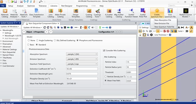
For the first case, if we want to use the simplistic tissue model and add fluorescence to it, we can simply select the layer of interest and apply settings in the properties window. Select Phosphors and Fluorescence then select Standard for all applications, Basic is only retained for legacy purposes. From there select the files used for Absorption, Emission and Quantum Yield, if needed you can create new files for each of these to suit your modeling needs. Also note that Mie scattering can also be used in conjunction with photoluminescence, this is useful when simulating droplets of a given size and their optical effect.
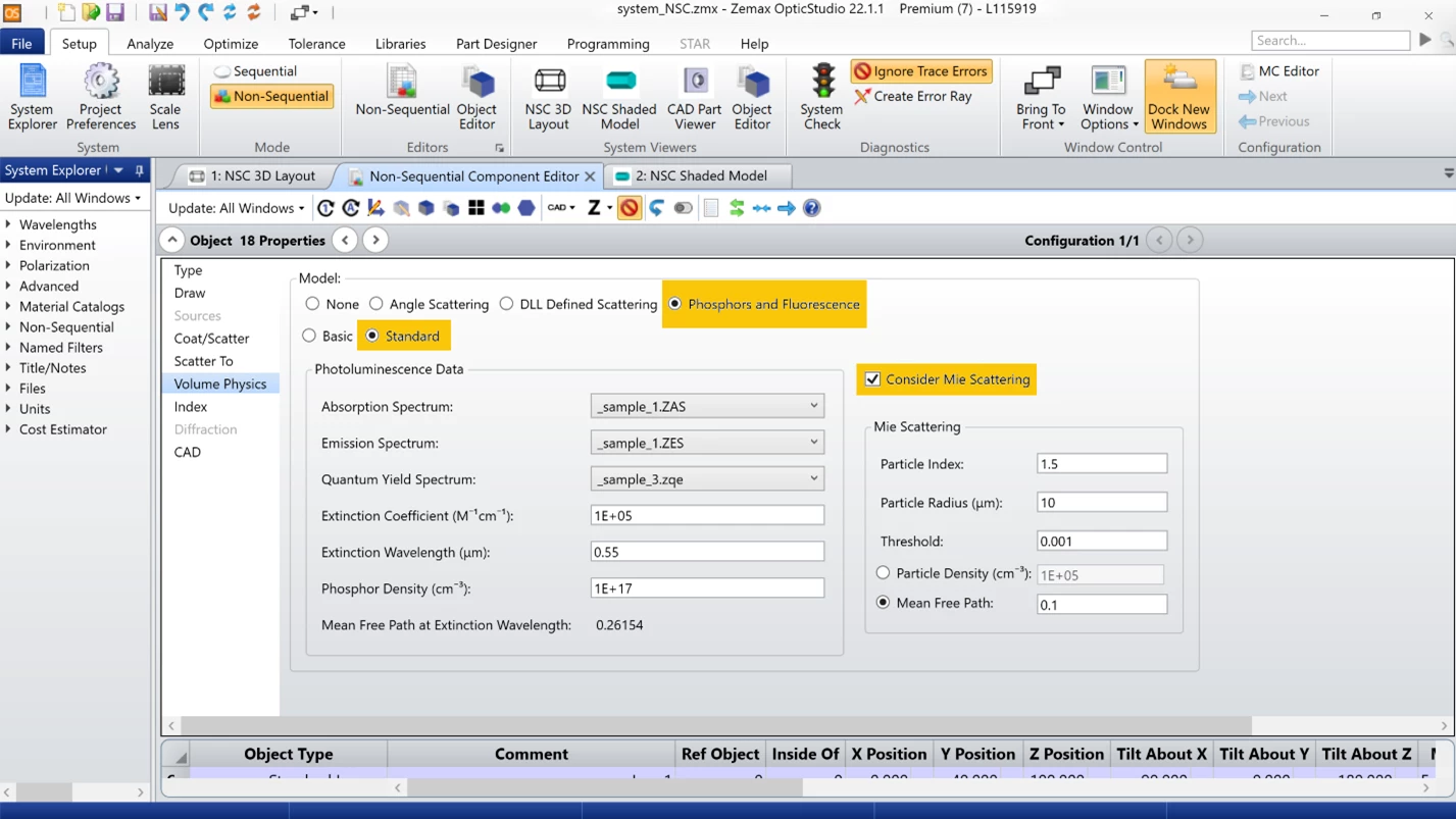
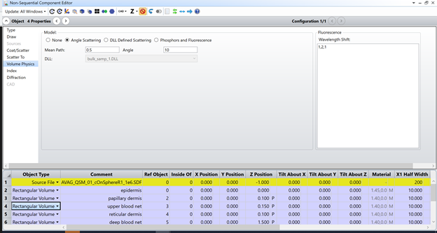
One note of caution here, Angle Scattering also has an option for Fluorescence, this is a wavelength shift from one defined wavelength to another. The numeric designation is referencing the wavelengths as they appear in the system explorer. This scattering option does not account for all the variables that the Phosphors and Fluorescence option can handle.
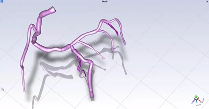
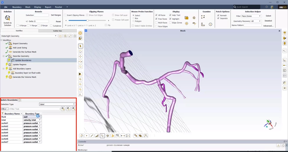
For our second case, let’s take this model further. It is possible to import a blood vein network created in Ansys Fluent as a CAD part and then assign Fluorescence and other optical properties to the object as would be done with any CAD model. The use of Fluent allows the user to simulate the turbulent mixing of the dye and blood, the chemical make up, viscosity, density and other physical properties are used allowing for the calculation of velocity of flow and the pressure at various points in the system, even allowing the user to analyze the system at different branches within a complex network. Fluent can also make use of the GRANTA MDS material database, or you can create your own materials and store them in the user defined database for future use. These options provide consistency across teams and reduce the time needed to input parameters for items that are often used. Leveraging these additional Ansys tools creates a far more realistic and wholistic model than can be achieved with OpticStudio alone. These considerations will also provide interesting inputs for OpticStudio to model the optical ramification of such a system.

Author: Steven LaCava Senior Application Engineer