Solved

Prescription Data explanation about Object space NA

  • 31 August 2022
  • 8 replies
  • 1239 views

Badge

Hi everyone,

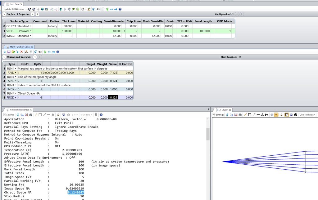
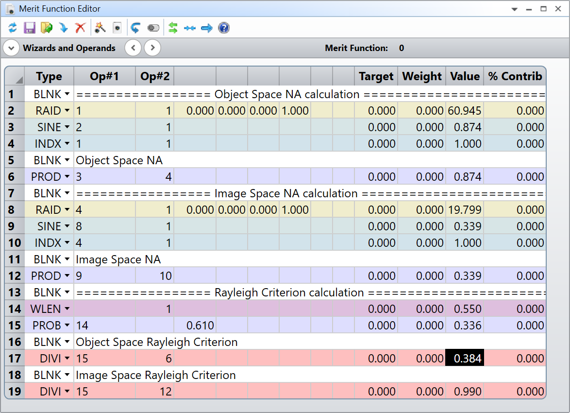
I got the Object Space NA of our system from Prescription Data, and try use it to calculate resolution with resolution formula (lambda/(2*N.A.)). However, it is off a lot. Does this object space NA in prescription data define differently from what we learn from Relay Criteria?

Thanks,

Xiaolei

icon

Best answer by David.Nguyen 7 September 2022, 12:22

View original

8 replies

Userlevel 7
Badge +2

Hi Xiaoleiwang,


The Object Space NA should be calculated as the refractive index at the desired wavelength times the sine of the marginal ray angle with the optical axis. As a sanity check, it can be computed in the Merit Function with the operands RAID, SINE, INDX, and PROD (see my screenshot below):

Can you tell us more about your system? There’s an article on the resolution in the Knowledgebase, perhaps it can be helpful.

Take care,


David

Badge

Hi David,

Thank you for your quick reply. The N.A. calculation is correct, I follow that. I am confused with the relation between Object Space NA and resolution of system. We have two stereomicroscope systems, the first one has Object Space NA 0.004, the second one has Object space NA 0.045. So if we use the resolution formula (lambda/2/N.A.), then the second one should has better resolution (high spatial frequency differential). However, the MFT gave me opposite results (see graphs below), which is the first one has high spatial frequency differential (better resolution). I don’t know what I miss here. Any information would be helpful!


Thanks,

Xiaolei

Userlevel 7
Badge +2

Hi Xiaolei,


I think I know what the problem could be. When you create an MTF analysis from OpticStudio, it is calculated at the image plane by default. This is indicated by the Surface setting of the analysis. Therefore, it should depend on your image-space NA (unless I’m mixing things up). Can you check what your image-space NA is and confirm whether this could be the issue?

Take care,


David

Badge

Hi David,

That’s right. The Image Space NA  of the first system is 0.06, while the second one is 0.04. So it make sense that the first system has better MTF than the second system. I used to work about confocal microscope, and the image resolution is determined by the objective NA, not image space NA. I think the stereo system is very different, and the image resolution is determined by the image space NA. Also, could you elaborate me how zemax calculate paraxial magnification and angular magnification? Thanks a lot! I really appreciate your insight and help!

Best,

Xiaolei

Userlevel 7
Badge +2

Hi Xiaolei,


The resolution is not necessarily determined by the image space NA. It depends what element limits the NA in your system. Sometimes, it can even be the illumination NA that matters. The issue with looking at the resolution in the image plane is that you have to factor in the magnification of your system. Let me show you in an example.

Consider the following 10X paraxial microscope:

The objective lens (on the left) has a focal length of 10 mm and a numerical aperture of 0.874. The tube lens (on the right) has a focal length of 100 mm and the image space NA is 0.177. The Rayleigh criterion using the object space NA is 0.384 um, and 1.894 using the image space NA. I’m showing the calculation in the Merit Function below together with a FFT PSF (at the image plane, this is analogous to MTF) showing the first zero occuring at about 1.894 (as predicted by Rayleigh):

Now, let’s look at what happens if we change the tube lens focal length to 50 mm:

As you can see, on the Object side, everything is still the same. But, as expected, the image side NA, and thus Rayleigh Criterion, changed. The new Rayleigh criterion on the image side is 0.990 um. Does that mean the system has a better resolution when compared to the 1.894 um derived with the 100-mm tube lens? Well, not really, and the reason is because the microscope is now 50X instead of 100X (50 mm / 10 mm = 5X). The image side PSF is roughly half the size (0.990 um vs 1.894 um) so two PSFs require half the distance to be discerned. However, translated in object plane, there’s a 2X change in the magnification (5X vs 10X). So, a distance in image side is twice larger in object side thereby canceling the gain in Rayleigh Criterion. That is why it is better to use the object-side NA, probably even for stereomicroscopes. You may want to try to model your microscope in the other direction, having the sample on the right. Also, I strongly suggest reading the article I was refering to before.

As for your last question about paraxial magnification. You can find more details in the Help file about the corresponding Merit Function operand. Here is an excerpt for:

  1. PMAG (paraxial magnification): 

    PMAG

    Paraxial magnification. This is the ratio of the paraxial chief ray height on the paraxial image surface to the object height at the wavelength defined by Wave. Only useful for finite conjugate systems. Note the paraxial image surface is used even if the system is not at paraxial focus.

  2.  AMAG (angular magnification):

    AMAG

    Angular magnification. This is the ratio of the image to object space paraxial chief ray angles at the wavelength defined by Wave. Not valid for non-paraxial systems.

Let me know if this helps.

Take care,


David


Badge

Hi David,

That’s right. When I worked on confocal microscope and epi-fluorescence microscope, I always use objective N.A. (object side N.A.) to calculate the image resolution, and use PSF with fluorescence beads to verify the image resolution. But for stereomicroscope, we use MTF to check the image resolution, and use USAF 1951 glass slide ( https://www.edmundoptics.com/f/1951-usaf-glass-slide-resolution-targets/12064/) to verify the image resolution. That’s why I am confused. I thought if MTF is better, then the image resolution should be better. But from the comparison of these two systems, the MFT better doesn’t mean the image resolution is better, right? Please correct me if I am wrong or miss something here. Thank you so much for your help!

Best regards,

Xiaolei

Userlevel 7
Badge +2

Hi Xiaolei,


My point earlier, and apologies for not making it clear, is that if you look at the image-side MTF, you need to account for the microscope magnification. Do the two microscopes have the same magnification?

Take care,


David

Badge

Hi David,

You are right, the two microscopes have different magnifications. The magnification of first one is 0.07 (Object Space NA 0.004, better MTF, WFNO: 8.03), while the magnification of the second one is 1.0(Object space NA 0.045, worse MTF, WFNO:11.46). My experiment test shows that the second one has high resolution than the first one. Could you tell me how Zemax calculate MTF? Also can I also calculate Field of View in zemax?

Thanks,

Xiaolei

Reply